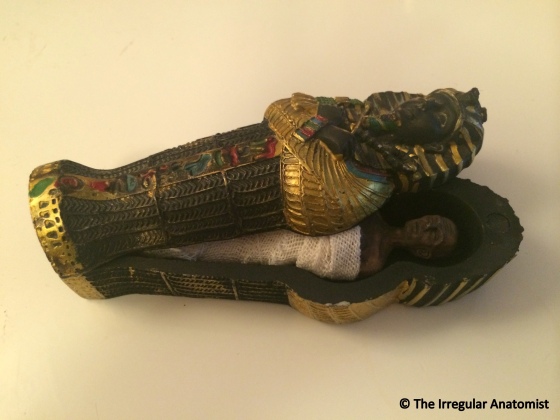
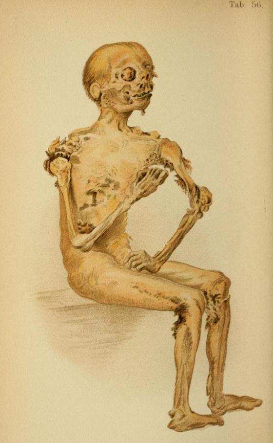
My mummified Egyptian in his sarcophagus
During a lovely, sunny break in Egypt this summer, I swore to myself I wouldn’t return home with typical, tacky, tourist tat. I failed. It’s just too easy to get carried away when shopping in Egypt. Initially wary of the market-place style haggling, it turned out to be one of the highlights of our holiday, spending some good times laughing and bantering with the local shop owners. It was on one of these occasions that I broke my tat-less promise. Shelves upon shelves of knick-knacks (including a lot of confusing penis-adorning Egyptian ornaments) I somehow managed to spot a gruesome gem. Imagine my deathly delight when I came across, not only a mummy’s sarcophagus, but one containing a creepy mummified corpse! The shopkeeper, clearly taken aback by my physical reaction to this morbid little mummy turned to my partner as asked, “She likes this kind of stuff?”. “Oh yeah,” he replied, “she loves the dead stuff!” Next came the customary ‘are you a bit weird?’ look that I’ve become accustomed to accept in such situations. The shopkeeper in a surprising ‘why didn’t you say so’ manner began to hunt high and low amongst the thousands upon thousands of goodness knows what that lined the shelves on the walls. “Here he is!” he exclaimed, “Anubis. The God of Death.”
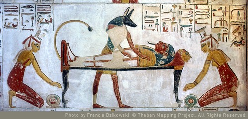
Anubis tends to the mummification process
One of the most iconic of the Ancient Egyptian gods, Anubis was known as guardian and protector of the deceased, their mummification and their journey through the underworld. A man with the head of a jackal, Anubis’ black appearance depicted death and fertility, representing rebirth and the afterlife. In the many statutes, artworks and archaeological finds in which he is depicted, he can be seen in various states of part jackal, part man but never as man in his entirety. Jackals and desert dogs would hunt at the edges of the Egyptian desert where the cemeteries and necropolis lay, like Anubis, watching over the dead.
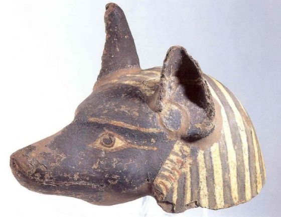
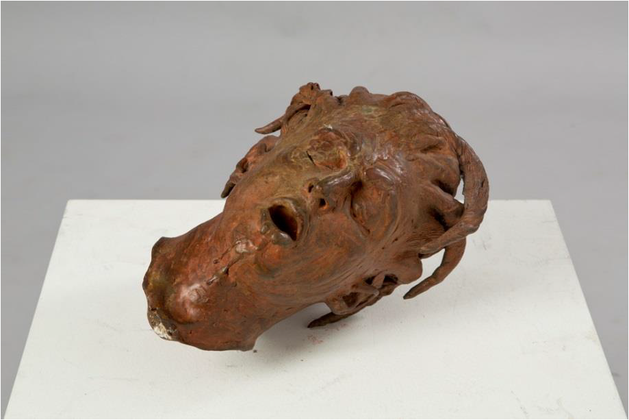
A mask of Anubis worn by priests during the mummification process. (Courtesy of Harrogate Borough Council, Museums and Arts Service)
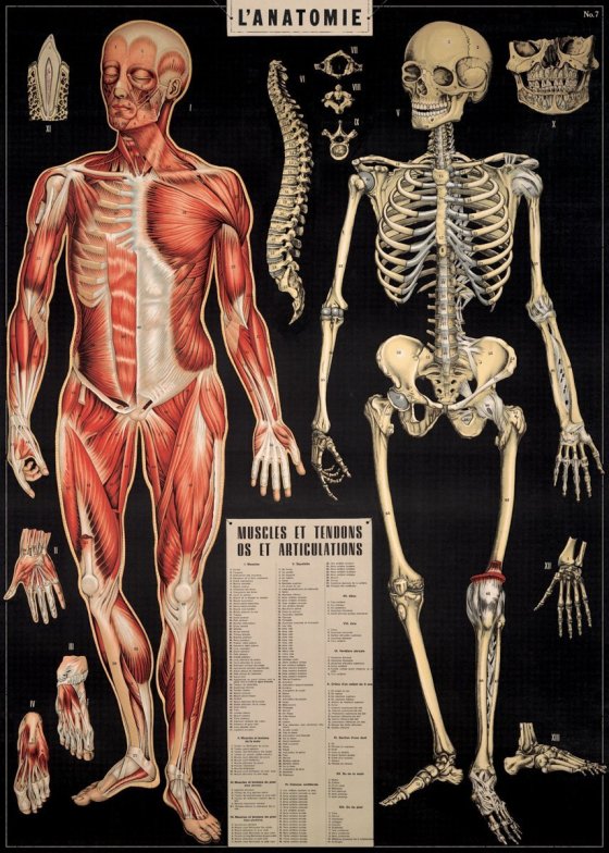
The Ancient Egyptians were famously known for the careful mummification of their dead whom they preserved with sweet-smelling plants and herbs. Anubis was thought to use his heightened sense of smell to sniff each mummified body and only allow the purest on to paradise. He would watch over the process, ensuring that the priests, who were known to wear masks of Anubis, conducted the mummification process to the utmost accuracy.
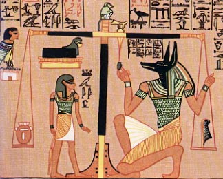
Anubis weigh a heart on the Scales of Justice
Anubis guided the souls of the dead through the underworld, testing an individual’s commitment to the gods and weighing their heart on the Scales of Justice. As Guardian of the Scales, Anubis would measure the weight of ones heart against the Feather of Ma’at which represented truth. If the heart was lighter than a feather, the deceased would be led into the underworld but if heavier, Anubis would feed the souls of sinners to Ammit the Destroyer; destructor of the souls of the corrupt and wicked.
So was my little Anubis worth taking home the tat for? According to the shopkeeper, I hope so; “Now you can have as much dead stuff as you want! Anubis will make you a mummy ever day. A hundred! You need him, he’ll always bring you dead stuff.” Sold. I’ll hold him to that.


 (
(